Services
Queries
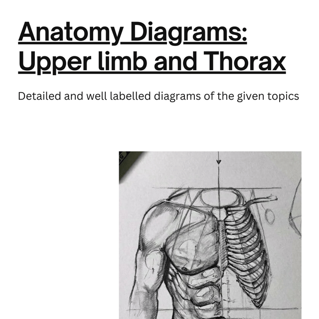
Upper limb and Thorax Anatomy Diagrams E-book
Frequently asked questions
What is histology?
Histology is the study of the microscopic structure of tissues — how cells are organised into epithelium, connective tissue, muscle, and nervous tissue. It is taught in the first year of MBBS and forms the base for pathology later, because you cannot recognise diseased tissue without first knowing what normal tissue looks like.
What are histology slides?
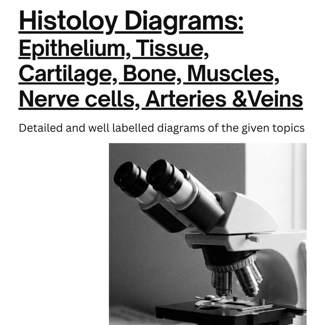
A histology slide is a very thin section of tissue, usually stained with haematoxylin and eosin (H&E), mounted on glass so it can be studied under a microscope. In MBBS practicals you are given such slides to identify, draw, and label, which is why students practice slide identification and diagram drawing together.
How to draw histology diagrams?
Start with a light pencil outline of the tissue's overall pattern, build up the cells layer by layer while referring to your slide or atlas, and finish with clean labelling and the stain used (for example, H&E). Keep the drawing large and simple, and redraw each diagram two or three times before exams — consistent practice matters far more than artistic talent.
How to prepare histology diagrams for MBBS 1st year?
Begin with your university's practical list, since most Indian universities test a similar core set — epithelium types, connective tissue, cartilage, bone, muscle, nerve, and organ sections like liver, kidney, and spleen. Draw each diagram once from the atlas and then again from memory, keeping labels such as lumen, capsule, and basement membrane consistent so you don't lose marks in spotters or practicals.
Is a handmade histology diagrams PDF enough for histology practicals?
Handmade diagrams are usually closer to what examiners expect than polished atlas plates, because they show the simplified, label-friendly version you are actually meant to reproduce in the answer sheet. Use a handmade set as your main drawing reference and keep a standard atlas alongside for finer detail — together they cover both practice and theory.
Where can I find a good histology diagrams PDF?
Look for diagram sets created by medical students who have recently cleared the same university pattern, because these focus on the slides that are actually asked. Student creators like the_artistic.medic share histology diagrams and accept direct requests, so you can get diagrams matched to your syllabus instead of hunting through scattered sources.
Do I need to prepare histopathology diagrams separately in 2nd year?
Yes, because pathology practicals move from normal tissues to diseased ones, so you will draw slides showing inflammation, granulation tissue, and tumours rather than healthy structures. Your first-year histology base makes this much easier — revise the normal appearance first, then learn what pathology adds or changes in it.
What is an anatomy diagram?
An anatomy diagram is a labelled drawing that shows body structures such as bones, muscles, vessels, nerves, and organs along with their spatial relationships. It is the standard way to present answers in anatomy theory papers, practicals, and vivas, which is why regular diagram practice is essential in the first year.
What are the most important anatomy diagrams for medical students?
For first year, give priority to the upper limb and thorax — muscle groups and compartments, the brachial plexus, bones and their attachments, and the heart with its chambers and blood flow. This is also why dedicated resources like the_artistic.medic's Upper Limb and Thorax Anatomy Diagrams e-book are so useful for first-year students.
Are anatomy diagrams PDF downloads enough, or should you draw them yourself?
PDFs are excellent for quick revision and last-minute reference, but reproducing a diagram from memory is what actually fixes the structure in your mind for spotters and vivas. The best method is to study a diagram from a PDF or e-book, draw it without looking, and then compare and correct your version.
Where can I find anatomy diagrams to label for practice?
Convert any diagram you already have into a labeling exercise — trace or print the outline, cover the labels, and fill them in against the clock. Practicing this way with unlabeled versions of key diagrams is one of the fastest ways to prepare for spotter rounds and viva questions.
What are pathology notes?
Pathology notes are condensed study material covering general and systemic pathology — disease definitions, etiology, pathogenesis, morphology, and complications — arranged in flowcharts, tables, and short points. Students use them for regular study, internal assessments, and especially for quick revision before university exams and profs.
How to make notes for pathology?
Make notes disease-wise rather than chapter-wise: one page per disease covering definition, etiology, pathogenesis, morphology, complications, and important lab findings. Use flowcharts for pathogenesis, comparison tables for similar diseases, and keep a separate one-page high-yield sheet per unit for the final week of revision.
Are pathology notes PDF files enough for university exams?
A good notes PDF covers most of what theory papers ask, but it works best alongside a standard textbook for topics you haven't understood and previous year question papers from your own university. Treat notes as a revision tool for what you have already understood, not as a first-time learning source.
How do I choose the best pathology notes for MBBS?
Pick notes that follow your university syllabus, use flowcharts and diagrams for morphology-heavy topics, and are short enough to finish in the last week before exams. Notes made by students who recently cleared the subject — such as the pathology notes the_artistic.medic shares — often match the exam pattern better than bulky material because they are written from the exam-taker's perspective.